Covalent organic networks are usually synthesized on noble metal surfaces. It is widely understood that these metals have strong catalytic abilities. However, it is of great interest the use of nonmetallic surfaces in these kind of reactions.
At the NanoPhysics Lab (CMF, Gipuzkoa) they’re studying one of these routes to obtain covalent molecular systems on non-metallic substrates. In particular they’ve managed to understand and improve the synthesis of nanoribbons on TiO2 surfaces. They show that highly reduced surfaces (in opposition to stoichiometric TiO2) increases the reaction yield and improved polymer length.
We did this picture to artistically illustrate the process under the close supervision of Dr. Celia Rogero.
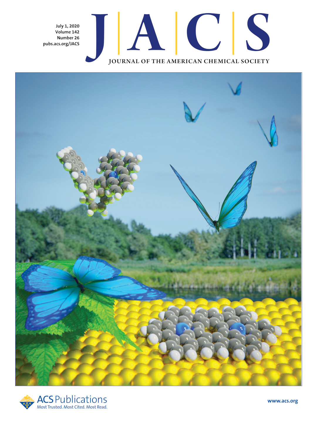
The level of control chemistry is reaching in the synthesis of graphene is mind-blowing. At the Department of Physics in Basel University, together with the University of Bern, Warwick and Lancaster, nitrogen-doped porous graphene nanoribbons (N-GNRs) were synthesized for the first time.
These N-GNRs are ladder-like molecules whose crystal lattice contains both periodic pores and a regular pattern of nitrogen atoms. And interestingly, these molecules don’t behave as conductors, as graphene does, but as semiconductors, making them very attractive in electronic applications.
We did this picture to illustrate the synthesis of the N-GNRs on request of Prof. E. Meyer and under the close supervision of Dr. Shi-Xia Liu.
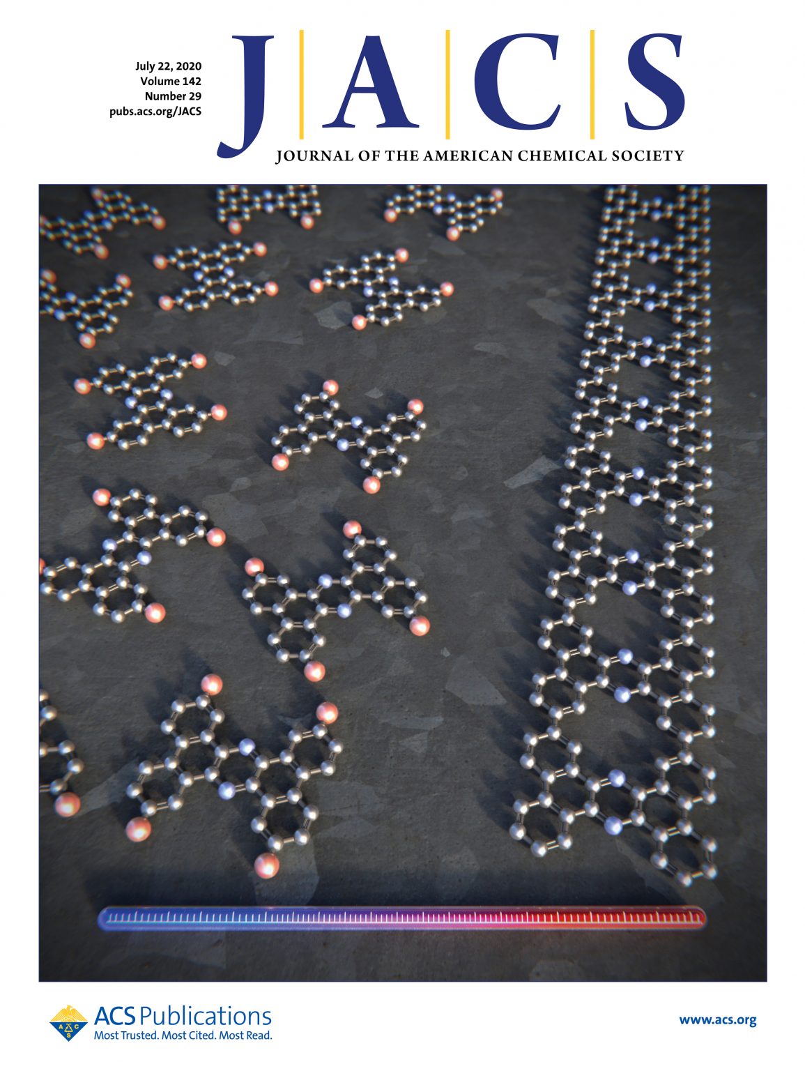
Circulene is a polycyclic aromatic hydrocarbon molecule composed by eight benzene rings. Because of geometric demands, the molecule adopts a saddle-shaped structure.This family of molecules, made of hexagonal and pentagonal rings are been studied for their promising applications in organic semiconductors, organic light-emitting diodes and liquid crystalline materials.
Prof. Shingo Ito et al. have just described the first example of a circulene bearing six hexagons and two pentagons which happens to have unique electronic structures, and intrinsic properties. In particular, they’ve proved this circulene to adopt a planar configuration.
This research, published in the Journal of American Chemical Society (JACS), was featured on the cover. The image was made under close supervision of Prof. Shigeky Kawai and Prof. Shingo Ito.
It’s not the first time we bring these people to our website, and there are several reasons for that. First of all, they keep hiring us to make cool images for them. But the main reason is that they are doing amazing work in the field of chemical sensors.
These time Dr. Juan Cabanillas-Gonzalez and Dr. Jose Sanchez Costa, both at IMDEA Nanociencia (Spain) bring us “A novel gas sensing mechanism exploiting lanthanide luminescence modulation upon NO2 adsorption”. To make a long story short, this is a crystal that glows beautifully when NO2 is around.
This has not only useful practical applications in the detection of NO2 market; it would make a beautiful luminescence displays without requiring expensive electronics. “But it also provides understanding of the nature and effects of NO2 interactions within the MOFs and the signal transduction mechanism.” You can read more about it in their recent article.
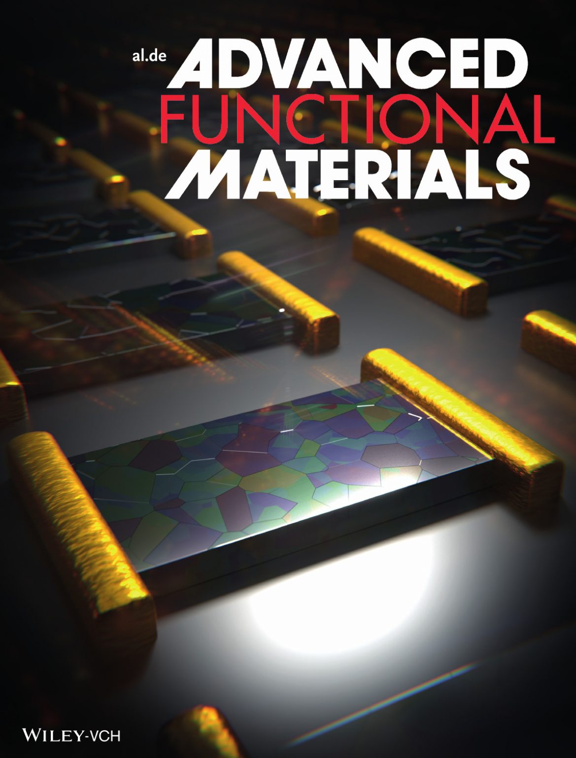
Despite his age, Dr. Mario Lanza has a long experience and deep knowledge on the physics and development of micro and nanoelectronic devices. In this recent article he an his coworkers discuss “the main challenges and potential solutions towards the fabrication of field effect transistors with 2D semiconducting channels”. In particular, there is a useful analysis on how this technology, now dealing with sizes that approach the interatomic distances, could be implemented in the current semiconductor industry.
This picture we did for him was featured on the cover of Advanced Functional Materials.
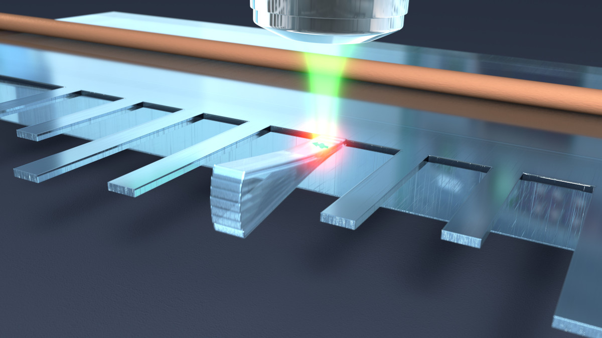
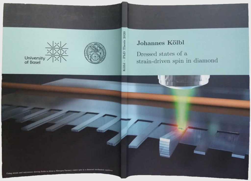
Quantum technologies building on solid-state spin systems, as those used in quantum computing and quantum internet, require unprecedented levels of stability. Even small fluctuations in ambient magnetic field messes up with the coherence time of these systems. So for these technologies to have any future we first have to learn how to stabilize and protect individual quantum ensembles. Doctor (congratulations on that) Johannes Koelbl at Basel University, has spent quite some time dealing with this challenge.
In this picture we made for him it is shown a Nitrogen-Vacancy center spin embedded in a diamond mechanical cantilever. As Dr. Koelbl explains: “To enhance its coherence properties, the spin is driven by both a time-varying strain field and microwave magnetic fields. The strain field is caused by harmonic oscillations of the cantilever, while the nearby wire serves as a near-field microwave source. Optical manipulation allows detecting the improved spin properties.”
He has chosen this picture to appear in the cover of his thesis manuscript.
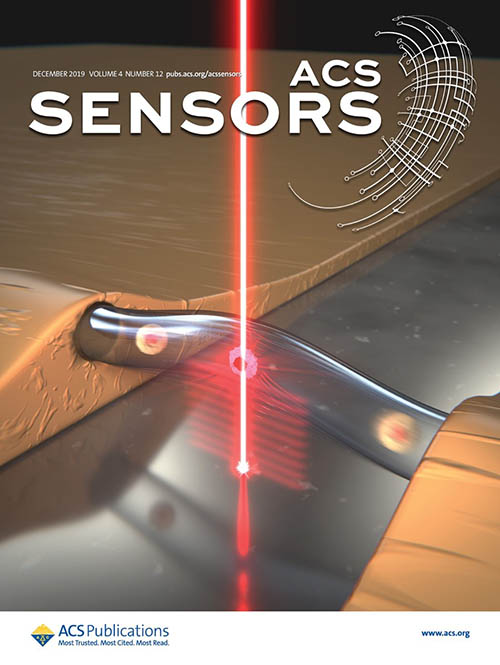
One of the last papers published by BioNanoMechanics Lab mixes biology, medicine, mechanics and optics. They have developed an efficient method to tell tumorigenic cells from healthy ones using mechanical and optical techniques. This group of researchers seems to be truly committed with the removal of the border between physics and biology.
The resonance frequency of an object ( ωf ) is amazingly sensitive to nearly everything, and in particular, to changes in mass of the object. Now imagine you built a very small glass capillary tube, you fill it with water with cells suspended in it. In this conditions, ωf will depend somehow in the mass of the cells. Now lets say, tumorigenic and healthy cells have different masses: there you have it! Your oscillating microcapillary tube is now a cancer detector.
Of course it is not that easy. To be sure they are measuring single cells and not clusters of cells or other suspended elements, they’ve added an optical probe, that produces, together with the mechanical data, a simultaneous optical measurement.
In summary, they’ve developed a novel, fast, efficient and beautifully ingenious way to detect tumorigenic cells.
This picture we did under the supervision of Montserrat Calleja and Alberto Martín was featured on the cover of ACS Sensors in December 2019.
We’ve had the fortune to work for researchers that study the drug delivery process. Ana Pizarro at IMDEA Nanociencia is focusing her efforts in the understanding of how and when to activate this drugs. In an article written for Chemistry A European Journal in 2017 she showed how to control in‐tumor drug activation via pH.
Innactive Ruthenium(II) arene complexes are innocuous and unable to interact with their molecular target. However, at a certain proton concentration this molecules are activated making it possible for them to bind to DNA.
Tumors happen to have a different pH than their environment making this complexes a possible option as drug switches.
I inexcusably forgot this work, considering the importance I give to medical related researchs. Her work was featured in the cover of the journal.
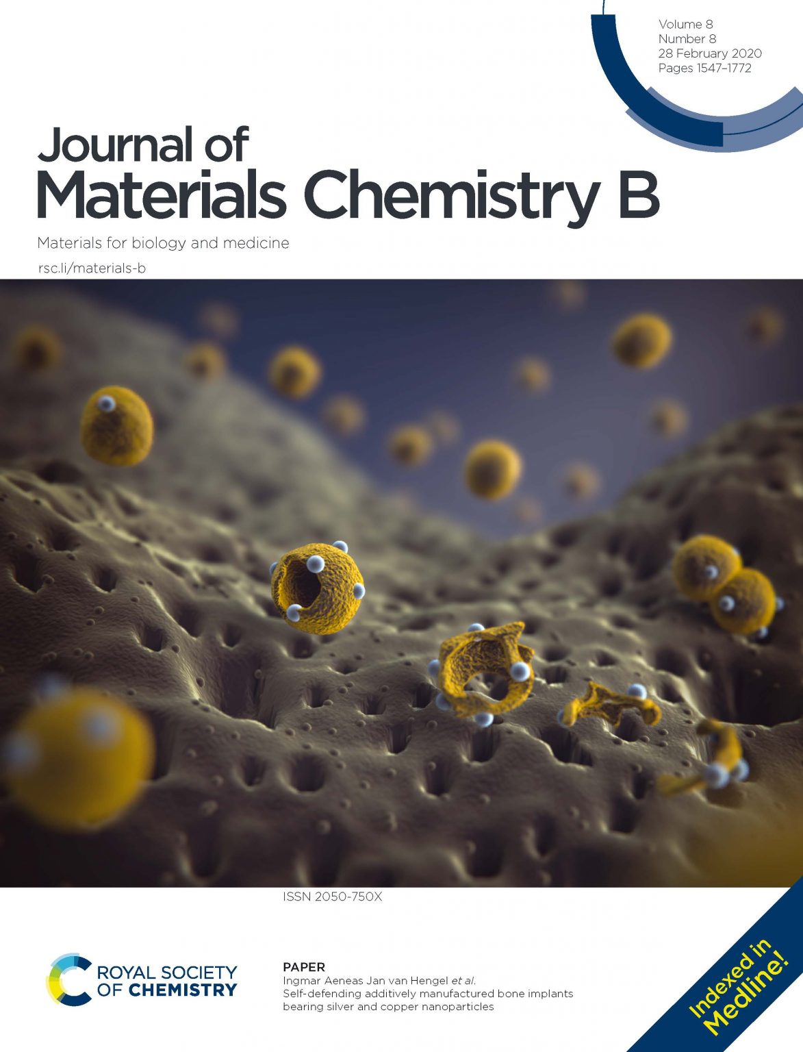
The manufacturing of bone implants involves a great deal of problems which are still to be solved. One of the most important challenges are implant-associated infections which make the development of implants with intrinsic antibacterial properties a pressing issue. This is precisely what they are trying to achieve at the Department of Biomechanical Engineering (TU Delft).
They’ve just studied the effect of both Ag and copper nanoparticles on TiO2 surfaces and its effectiveness as antibacterial and osteoconductive biomaterials. In fact they’ve observed that these materials “have a strong antibacterial behavior against both planktonic and adherent bacteria in vitro conditions.”
These results have been published in the Journal of Materials Chemistry B and have been featured on the cover. We designed the picture under the supervision of Ingmar A. J. van Hengel, first author of the paper.
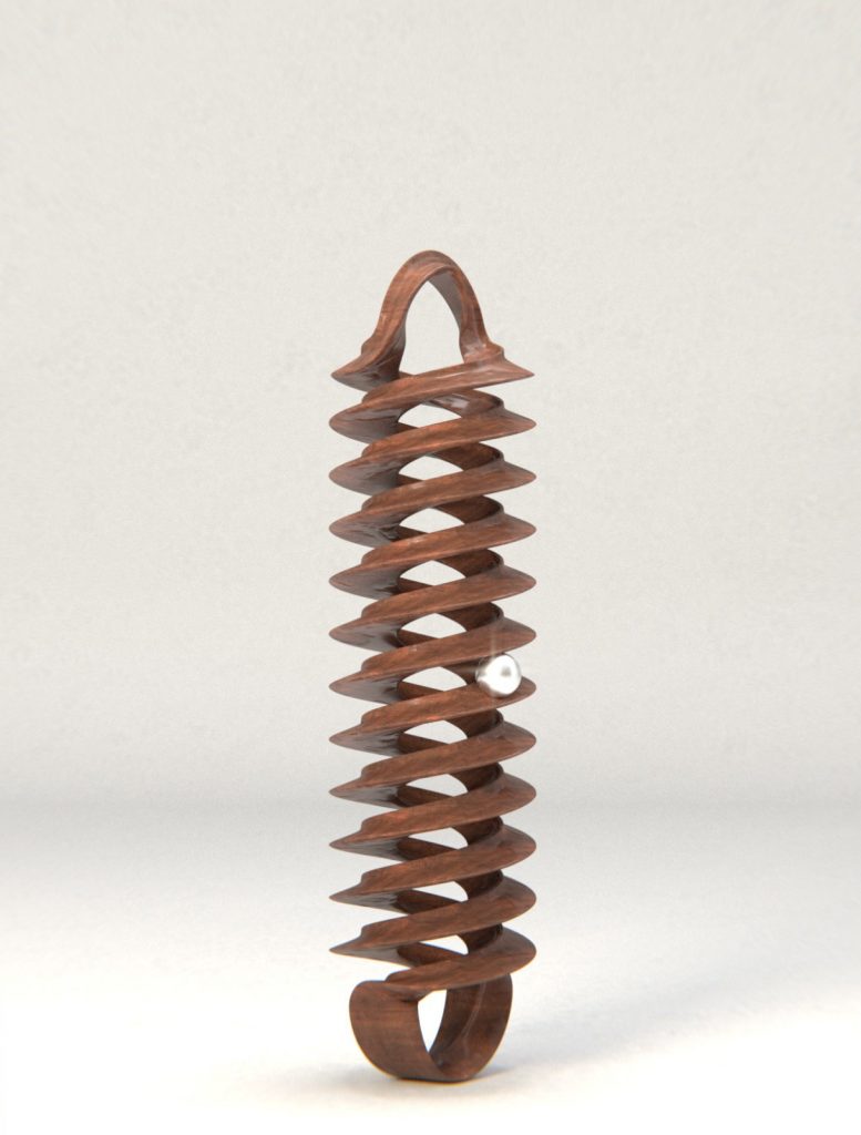
We, as human’s, are pretty familiar with the influenza A virus, so it is confusing to know how much there is still to learn about it. And researchers from CSIC have just reduced our ignorance about it a little bit more. Together with researchers from Stockholm University, CNRS, Université Paris Diderot and Institut Pasteur, and using cryoelectron microscopy, they’ve unveiled the transcription mechanism of this virus. This is important, among other reasons, to understand why this virus is so successful. And its been published in Nature Microbiology.
The molecules responsible for transcription are the ribonucleoproteins (RNPs). This RNPs which are extremely flexible, adopt a double helical conformation. In this configuration, the RNA, attached to the RNPs, slides in a sort of worm drive fashion. This process can be seen in the video we made for them as supplementary information for their paper. As put by the researchers, “the flexibility of the viral RNPs is key and explains how the virus is able to create a big amount of proteins from a limited number of genes”.
We also attempted the cover of Nature with this picture.
I’ve never whined about not getting a cover, but there is always a first. I’ve had the privilege to follow this research for about two years, thanks to Jaime Martín-Benito, so I can’t but feel it as something personal. The discovery is amazingly important and the picture is really beautiful (idea of Jaime Martín-Benito, corresponding author of the paper). And it deserves to be shown! So here it is for your enjoyment.